Histology core
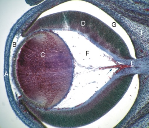 The HISTOLOGY portion of the Imaging and Histopathology Core is responsible for the processing, embedding, and sectioning of tissue samples. This includes samples processed for cryopreservation, paraffin, and Epon or other plastic embedding. A wide variety of ocular tissue samples from many different species are routinely processed.
The histology personnel produce professional quality slides for imaging to be used in research analysis, posters, presentations, publications and grants. Investigators are encouraged to contact the module manager prior to the conception of any histology based experiments. During consultation the module manager will discuss the most appropriate means to process your samples depending on your specific needs and desired results. As with all Core supported facilities, NEI R01 funded PI's have first access to the services provided. Most of the PI's in the WSU Vision Core utilize this module.
How to utilize
- Contact the module manager prior to beginning projects
- Complete the Imaging and Histopathology work order
- Provide the module manager with any special supplies needed to complete the project
Major instrumentation
- Microm HM 520 cryostat
- Microm HM 525 cryostat
- Histopro 150 H paraffin processor
- Leica RM 2235 paraffin microtom
- Reichert ultracut S ultratome
- HP Designjet T739 eprinter
Pricing information
- Utilizing Module Manager for training and non-routine consultation: $30.00/hour
- Cryosectioning Usage ($10/hr)
- Paraffin processing and sectioning (routine H and E) ($10/block)
- Poster printing ($65/poster; additional $20 for mini poster)
Please complete the work order and submit to core manager.
Sharon McClellan
Module Manager
Department of Ophthalmology, Visual and Anatomical Sciences
540 E. Canfield Avenue
7341 Scott Hall
Phone: 313-577-1074

Susmit Suvas. Ph.D.
Module Director
Professor - Department of Ophthalmology, Visual and Anatomical Sciences
Wayne State University School of Medicine
540 East Canfield Avenue
Detroit, MI 48201
Phone: 313-577-9820