Imaging core
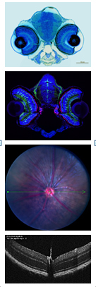
 The IMAGING portion of the Imaging and Histopathology Core has been a part of Core services for over 30 years. It provides user access to and training in the correct use of the SP8 scanning Laser confocal microscope, fluorescence microscopy, brightfield microscopy, slit lamp imaging, full color retinal fundus imaging, fluorescein angiography, and optical coherence tomography (OCT). Visual functional analysis instrumentation includes an ERG viewer and an optokinetics system. Users are required to be trained on the core's microscopes and other instrumentation and they can also be instructed on the proper use of any instruments that they have in their own labs. Additionally, the module manager maintains the day to day function of all the instruments including the Leica SP8 laser scanning confocal microscope and the Zeiss AxioImager, two of the most integral and heavily used instruments housed in the Core lab facilities. Image analysis services and training are also provided by this module. Investigators are encouraged to contact the module manager prior to the initiation of any microscopy based experiments. We will provide a review of the experimental design; suggest the correct selection of stains, dyes, or fluorophores as well as which microscope will best suit specific needs. These steps are all essential in obtaining consistent publication quality data. As with all Core supported facilities, NEI R01 funded PI's have first access to training and use of the equipment. Most, if not all PI's in the WSU Vision Core utilize this module.
Major instrumentation
- Cerebral mechanics Inc. Mouse OptoMotry system (OKT)
- Leica SP8 laser scanning confocal microscope
- Leica MZ10F Fluorescent stereomicroscope
- Leica DM4000b Brightfield microscope with full color digital camera system
- Ocuscience ERG Viewer
- Phoenix Micron V Digital Retinal Imager with OCT II
- Topcon Sl7E Slit lamp
- Viewlight 8U portable Slit lamp
- Zeiss AxioImager with apotome light sectioning attachment
- Leica Thunder Imaging System
Pricing information
- Utilizing Module Manager for digital analysis, training, repairs, non-routine consultation: $30.00/hour
- Leica SP8 Confocal Microscope: $45.00/hour
- Zeiss AxioImager: $15.00/hour
- Leica MZ10F fluorescent stereomicroscope with camera $10.00/hour
- Phoenix Micron V: $20.00/hour
- Leica Thunder Imaging System: $25.00/hour
Please complete the work order and submit to core manager.

Muhammed Farooq Abdul Shukkur
Module Manager
Department of Ophthalmology, Visual and Anatomical Sciences
540 E. Canfield Avenue
7341 Scott Hall
Phone: 313-577-1074

Susmit Suvas. Ph.D.
Module Director
Professor - Department of Ophthalmology, Visual and Anatomical Sciences
Wayne State University School of Medicine
540 East Canfield Avenue
Detroit, MI 48201
Phone: 313-577-9820